facs buffer flow cytometry
Resuspend stained cells in an appropriate volume of Flow Cytometry Staining Buffer. When gating on cell populations the light scatter profiles of the cells on the flow cytometer will change considerably after permeabilization.

Spillover Coefficient In Cytometer Flow Cytometry Experiments Molecular
Resuspend cells in 100 μL of Flow Cytometry Staining Buffer Catalog FC001.

. Antibodies should be prepared in permeabilization buffer to ensure the cells remain permeable. A washing step is not necessary. Flow Cytometry Panel Design SupportWork with one of our technical sales specialists to discuss your experimental needs and guide you through the process.
To adjust flow cytometer settings for PI add 5 - 10 μL of PI staining solution to a. Resuspend at 2 x 10 6 cells in 1 ml ICE COLD BUFFER. Stain cells using 05 ml HoechstPY staining solution.
The sample should be kept in the dark. BD Biosciences provides flow cytometers reagents tools and a wide range of services to support the work of researchers and clinicians who understand disease and improve care. Wash cells by centrifugation eg.
Cells are usually stained in polystyrene round bottom 12 x 75 mm 2 Falcon tubes. 1- Use CaMg2 free PBS. Flow Cytometry Support CenterFind technical support recommendations for your flow cytometry workflows including tips for experimental setup and in-depth troubleshooting help.
The samples should be resuspended in Cell Staining Buffer. Analyze samples by flow cytometer. However they can be stained in any container for which you have an.
General procedure for flow cytometry using a conjugated primary antibody. Harvest wash the cells and adjust cell suspension to a concentration of 1-5 x 10 6 cellsmL in ice-cold PBS 10 FCS 1 sodium azide. Our comprehensive reagent portfolio includes clinical diagnostic testing kits kits for innovative new approaches to clinical research and single-color reagents for.
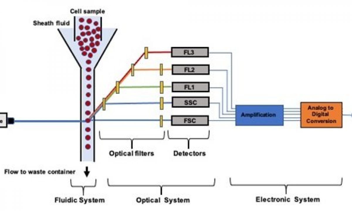
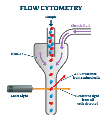
Flow cytometry has been extensively exploited in immunology hematology and oncology to define cell populations via intrinsic scatter properties cell surface antigen expression and other fluorescence parameters 1-3Our insights into blood lineage development and disease are a result to a significant degree of the continuous refinement of. Incubate 20 min at room temperature and analyze the. 200 x g 5 min 4C in protein-free buffer such as Phosphate Buffered Saline without Ca 2 or Mg 2 PBS.
Perform fluorescence activated cell sorting FACS or flow cytometric analysis. A debris free single cell suspension will have lower auto-fluorescence and flow smoothly through the. BD Biosciences flow cytometry reagents truly reflect our scientific leadership in flow cytometry innovation and our 45 years of dedication to providing high-quality products.
Add 200 µL Flow Cytometry Staining Buffer and centrifuge at 600 x g for 4-5 minutes. Here are 5 ingredients to consider for your FACS buffer. Absence of these ions reduces cation-dependent cell to cell adhesion and prevents clumping.
Flow cytometry FACS staining protocol Cell surface staining Harvest wash the cells single cell suspension and adjust cell number to a concentration of 1-5x10⁶ cellsml in ice cold FACS Buffer PBS 05-1 BSA or 5-10 FBS 01 NaN3 sodium azide. Detecting intracellular antigens requires cell permeabilization before staining. Optional Repeat step 1.
Cell number will effect staining qualityOptional. If you are unable to immediately read your samples on a cytometer keep them shielded from light and in a refrigerator set at 4-8C. Wash cells with 5 ml FACS buffer twice by centrifuging 5 min at 200 g.
If MOG-IgG1 cell based flow cytometry FACS assay is positive at screening dilution the MOG-IgG1 flow cytometry titer assay is performed at an additional chargeUnpublished Mayo method PDF Report Indicates whether the report includes an additional document with charts images or other enriched information. Use pre-coated or silanized polypropylene tubes to minimize sticking.
Microfluidic Flow Cytometry Principles And Commercial Review Ufluidix

Flow Cytometry Protocols
Ebioscience Flow Cytometry Staining Buffer

Flow Cytometry Basics Flow Cytometry Miltenyi Biotec Technologies Macs Handbook Resources Miltenyi Biotec Usa
Flow Cytometry Perm Buffer 10x Pf00011 C Proteintech

What Are The Uses Of Flow Cytometry Enzo Life Sciences
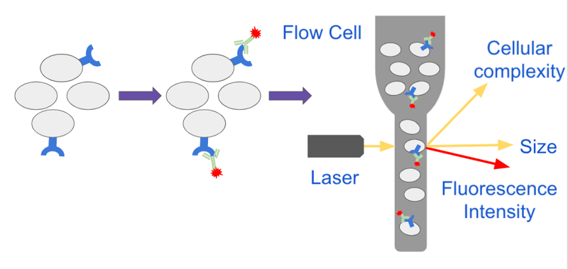
Analyzing Single Cells With Flow Cytometry

Fundamentals Of Flow Cytometry Aat Bioquest

What Is Flow Cytometry Technology Networks
Flow Cytometry And Cell Sorting Core Utmb
Intro To Flow Temerty Faculty Of Medicine Flow Cytometry Facility
Flow Cytometry Guide Creative Diagnostics

Flow Cytometry Facs Protocols Sino Biological
Popular Antibodies For Flow Cytometry Proteintech Group

Immunophenotyping Kits For Flow Cytometry Thermo Fisher Scientific Es
Flow Cytometry Control And Standardization Beads
Quantitative Flow Cytometry Measurements Nist

Ldl Uptake Assay Kit Flow Cytometry Ab236208 Abcam

Dapi Staining And Flow Cytometry Analysis Of Ht 29 Cells Treated With Download Scientific Diagram